Tag: Hospital Sant Pau

  • DIAGNOSIS SOFTWARE

    DIAGNOSIS SOFTWARE

    Project Description

    For computation of motion estimation in tagged-MRI, the interactive and augmented modelling group  has developed the Harmonic Phase Flow (HPF) plugin. HPF is a variational approach that works in the Gabor domain, ensuring optical flow invariance as well as motion tracking robustness. Additionally, HPF computes wall deformation without overestimating motion at injured areas.


    CLIENT

    Hospital Sant Pau

    Tagged MRI

    Since the development of Tagged MRI, in the late 80’s, many analysis techniques have been developed for motion extraction. Unfortunately, tagging studies are still not widely used in the clinical routine for two reasons:

    • There is a lack of reference values for new data derived from such imaging modality (stress, strain, torsion)
    • Commercial software capable of dealing efficiently with the great amount of data provided by the tagging studies is scarce
    Clinical tool

    Osirix is a popular opensource DICOM viewer for the Mac. It is actively developed by Pixmeo, Switzerland. It has become popular in the last years, since it integrates a user friendly interface for medical imaging visualisation with powerfull plugin system that extends its functionaly, enabling advanced image processing techniques

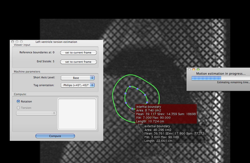
    The Sant Pau hospital uses OsiriX as the preferred tool for TMR sequence viewing. Because of this, it is interested in the development of a plug-in for the extraction and validation of myocardial torsion from their TMR sequences. We have previously built a prototype in Matlab for this purpose. However, communication between Matlab and OsiriX is slow and memory intensive. Therefore we have decided to port existing code into Objective C.

    Technologies

    Cocoa

    An object oriented framework for Mac OS X. Communication between the graphical user interface elements and inner data requires the design of cocoa objects with the Objective-C language.

    VTK

    The Visualization Toolkit (VTK) manipulates data, reconstruct multiplanar slices, performs volume and surface rendering with 2D or 3D texturing.

    Core-plot

    Core-plot is a recent plotting library for MAC OSX and IOS. It allows for rich visualization of the torsion data.

    Objective C

    A dynamic, object-oriented language, based on a set of extensions of ANSI C. It is the language of choice for Cocoa development.

    ITK

    The Insight Toolkit (ITK) is an open-source library for image registration and segmentation written in C++. Its foundation is the VNL (numerics) library for matrix algebra, which we also use in our own code.

    FFTW

    The Fastest Fourier Transform in the West (FFTW) is an implementation of the discrete Fourier transform (DFT) that adapts to the hardware in order to maximize performance.

    OpenGL

    The graphics API choice for both 2d and 3d rendering. At large sequence lengths, it is beneficial for fast visualisation operations.

    DCMTK

    Papyrus toolkit and DICOM Offis (DCMTK) handle specific details of the DICOM image format used by the TMR sequences.

    Alglib

    Alglib is a cross-platform numerical analysis and data processing library used for interpolation of the rotation of two LV views with different -end-diastole lengths

    Our solution

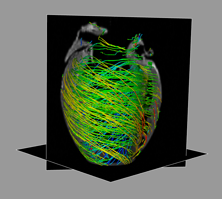
    We implemented an in-house fully automated method for tissue deformation estimation in tagged magnetic resonance images (TMRI). We call this schema the Harmonic Phase Flow. The method consists of two main stages, namely Gabor Filter Bank computation and sequence filtering.

    Gabor Filter Bank computation

    We built two Gabor filter banks for each tag orientation. Both banks are fine-tuned  for each left ventricle level. This allows for an optimally filtering of  complex images. This is crutial, since  the complex phase   remains constant along the cardiac cycle. This  brightness constancy condition is required by classical optical flow (OF) methods.

    Sequence Filtering

    Both filtered sequences belonging to a variational formulation schema  are used in a to obtain a dense continuous deformation map. We consider this as Harmonic Phase Flow.

    This method has been used to determine reference values of ventricular torsion (VT) in a set of 40 healthy volunteers. The results encourage the use of ventricular torsion as a useful parameter for ventricular function assessment in clinical routine.

    Implementation

    Due to the complexity of the original implementation, we broke down the process into several tasks.

    OsiriX platform acquaintance

    To translate Matlab code into native Objective C, we looked at other existing plug-in implementations, such as Brian Jensen’s NMSegmentation plugin from the Technische Universität München. In order to familiarise with the OsiriX plug-in SDK, we developed an initial version which only used a fixed set of points as the input LV. After this, we evolved these points in time using a set of pre-calculated HPF motion matrices. We consider the ventricular torsion as the difference in rotation at two different Short Axis views of the same sequence. To validate our results, we measured the absolute error between the global mean VT of our prototype and Matlab. Additionally, we ensured that the plot curves followed the expected tendency in both cases.

    HPF computation

    HPF is a regularised spectral optical flow, under a variational framework that uses the maximum response of a set of Gabor filters banks. This flow characterises the ventricular torsion in healthy subjects.  In step 1, we detect the harmonic peak which we use for building the Gabor filter banks (step 2). The combination of the response of banks produces a complex image whose phase is related to tissue motion (module 3). Finally, the we track the amplitude of the maximum response for building the displacement map.

    Get the plugin now!

    DISCLAIMER

    This tool is designed for clinical research only. Harmonic Phase Flow is not a medical device, and is not certified by any official organism, including the Federal Drug Agency (FDA) or the European Medicines Agency .

    Licence: GNU public licence v3.

    This program is free software: you can redistribute it and/or modify it under the terms of the GNU General Public License as published by the Free Software Foundation, either version 3 of the License, or (at your option) any later version.

    This program is distributed in the hope that it will be useful, but WITHOUT ANY WARRANTY; without even the implied warranty of MERCHANTABILITY or FITNESS FOR A PARTICULAR PURPOSE. See the GNU General Public License for more details.

    You should have received a copy of the GNU General Public License along with this program. If not, see <http://www.gnu.org/licenses/>.

    Citation

    Albert Andaluz, Francesc Carreras, Cristina Santa Marta, & Debora Gil. (2012). Myocardial torsion estimation with Tagged-MRI in the OsiriX platform. In Wiro Niessen (Erasmus MC) and Marc Modat (UCL) (Ed.), ISBI Workshop on Open Source Medical Image Analysis software. IEEE.

    Sample sequence

    Base

    Mid

    Apex

    Documentation

  • CARDIAC IMAGING ANALYSIS

    CARDIAC IMAGING ANALYSIS

    Project Description

    Miocardia is a solution pack which provides a unique comparative setting of multiple cardiac imaging modalities for the modeling and representation of the Left Ventricle Functionality. Miocardia is meant to be Computer Vision software capable to represent the anatomical and functional structure of a ventricle on 3D. It extracts information from different kind of image modalities of the patient’s heart to build the 3D representation. Deep understanding of myocardial structure of the heart would unravel crucial knowledge for clinical and medical procedures. Although the muscular architecture of the heart has been debated by countless researchers, the controversy is still alive. Diffusion Tensor MRI, DT-MRI, is a unique imaging technique for computational validation of the muscular structure of the heart. By the complex arrangement of my- ocites, existing techniques can not provide comprehensive descriptions of the global muscular architecture. We introduce a multiresolution reconstruction technique based on DT-MRI streamlining for simplified global myocardial model generation. Our reconstructions can restore the most complex myocardial structures and indicate a global helical organization.


    PARTNERS

    Hospital Sant Pau

    FUNDING

    TIN2009-13618


    Fiber Reconstruction

    Deep understanding of myocardial structure of the heart would unravel crucial knowledge for clinical and medical procedures. Although the muscular architecture of the heart has been debated by countless researchers, the controversy is still alive. Diffusion Tensor MRI, DT-MRI, is a unique imaging technique for computational validation of the muscular structure of the heart. By the complex arrangement of myocites, existing techniques can not provide comprehensive descriptions of the global muscular architecture.We introduce a multiresolution reconstruction technique based on DT-MRI streamlining for simplified global myocardial model generation. Our reconstructions can restore the most complex myocardial structures and indicate a global helical organization.

    Left Ventricle Functionality

    Most cardiovascular diseases impair the normal motion of the left ventricle (LV). Recent improvements in tomographic imaging devices such as MRI or CT, have led to the creation of 3D computer models that show the dynamics of the LV. Since the mechanical properties of any muscle are determined by the arrangement of their fibres in space, a model that aims to realistically simulate the behavior of this tissue must take into account the spatial distribution of the fibres. In the case of the myocardium it plays a crucial role for the understading of the electromechanical activation sequence in normal and pathologic conditions and the mechanisms of the ventricular resynchronization therapy (pacemaker implantation).

  • MEDIAL SURFACE COMPUTATION

    MEDIAL SURFACE COMPUTATION

    Project Description

    Medial branching is usually associated to perturbations of the object bound- ary rather than to its main geometric features. Such instability of the medial surface is a main obstacle for its confident application in shape recognition and description. Medial branches correspond to singularities of the medial surface and, thus, they are problematic for existing morphological and energy-based al- gorithms. In this paper, we use algebraic geometry concepts in an energy-based approach to compute a medial surface presenting a stable branching topology. We also present an efficient GPU-CPU implementation using standard image processing tools. The higher performance of our method in terms of compu- tational cost and medial surface quality is shown on a home-made synthetic database. Finally, we present some results on a medical imaging application for localization of abdominal pathologies.


    PARTNERS

    Hospital Sant Pau, AlmaITSystems, Universitat Pompeu Fabra, Hospital Clínic

    FUNDING

    TIN2012-33116

  • RHEUMATOID ARTHRITIS

    RHEUMATOID ARTHRITIS

    Two stages are involved in this task. Firstly, the joint detection, afterwards, the joint characterization. Unlike the little existing previous work, this contribution clearly separates those two stages and sets the foundations of a modular assessment system focusing on the characterization stage. A hand joint dataset is created and an accurate data analysis is achieved in order to identify relevant features.

    Since the sclerosis and the lower bone were decided to be the most important features, different computer vision techniques were used in order to develop a detector system for both of them. Joint space width measures are provided and their correlation with Sharp-Van der Heijde are verified.

    Project Description

    Rheumatoid arthritis (RA) is an autoimmune inflammatory type of arthritis which mainly affects hands on its first stages. Though it is a chronic disease and there is no cure for it, treatments require an accurate assessment of illness evolution.Such assessment is based on evaluation of hand X-ray images by using one of the several available semi-quantitative methods.This task requires highly trained medical personnel. That is why the automation of the assessment would allow professionals to save time and effort.


    CLIENT

    Hospital Sant Pau

  • XYLEM 3D RECONSTRUCITON

    XYLEM 3D RECONSTRUCITON

    Extraction of the xylem network characteristics (conduits and membranes defining their connectivity) is achieved using Micro-CT series.

    A main challenge is preserving small conduits (of the order of microns), as well as, thin separating membranae connecting conduits in highly noisy images. A structure preserving smoothing (SPD) developped in our lab is applied to filter texture while preserving consistent biological structures. After such smoothing, simple morphologic operators reconstruct the 3D topology of the Xylem network.

    Project Description

    Plants transport water from root to leaves through a network of tubular conduits call Xylem. The conduits size and connectivity determines the plants hydraulic properties. Micro-CT series have recently arisen as a powerful imaging modality for extracting the xylem network.

    The project consists of the development of a computer vision system for 3D modeling and recontruction of plant xylem network in order to determine plants resistance to extreme climatic conditions. The 3D recontruction task of Xylem net is done using Micro-CT image processing. Extraction of  the xylem  network characteristics (conduits and  membranes defining their connectivity) is  achieved using Micro-CT series.


    CLIENT

    Hospital Sant Pau