
Project Description
iVENDIS: Intelligent In-vivo Endoscopy Navigation, Diagnosis and Intervention Support systems.
One of the main limitations of endoscopic procedures is the impossibility to get accurate 3D measures and location of the injured tissue which may lead to the repetition of some interventions with an additional cost for the healthcare system. Another limitation is the lack of objective tissue characterization to allow in-vivo diagnosis, delaying decision taking and patient treatment beginning. Therefore, clinicians are in urgent need of systems able to provide on-line 3D measurements, 3D positioning and objective tissue characterization tools during the endoscopic intervention. iVENDIS aims at on-line extraction of physiological data from endoscopy interventional videos for building up better lesion detection, diagnosis and treatment planning systems.
iVENDIS is composed by three main products: SENSA, 3DEND and SAIPHIC.
Machine Vision group is also involved in this project.
CLINICAL PARTNERS
Hospital Clínic de Barcelona, Hospital de Bellvitge
FUNDING
Fundació La Marató de TV3, TIN2012-33116, 2014PROD00065, DPI2015-65286-R
SENSA
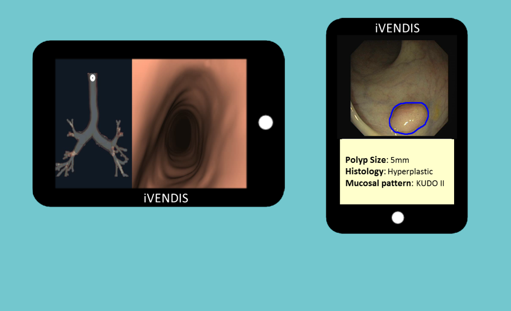
A system for Endoscopic Stenosis Assessment (SENSA) for real- time segmentation of tracheal main anatomical structures in videobronchoscopy to provide accurate standardized measurement of tracheal obstructions.
3DEND
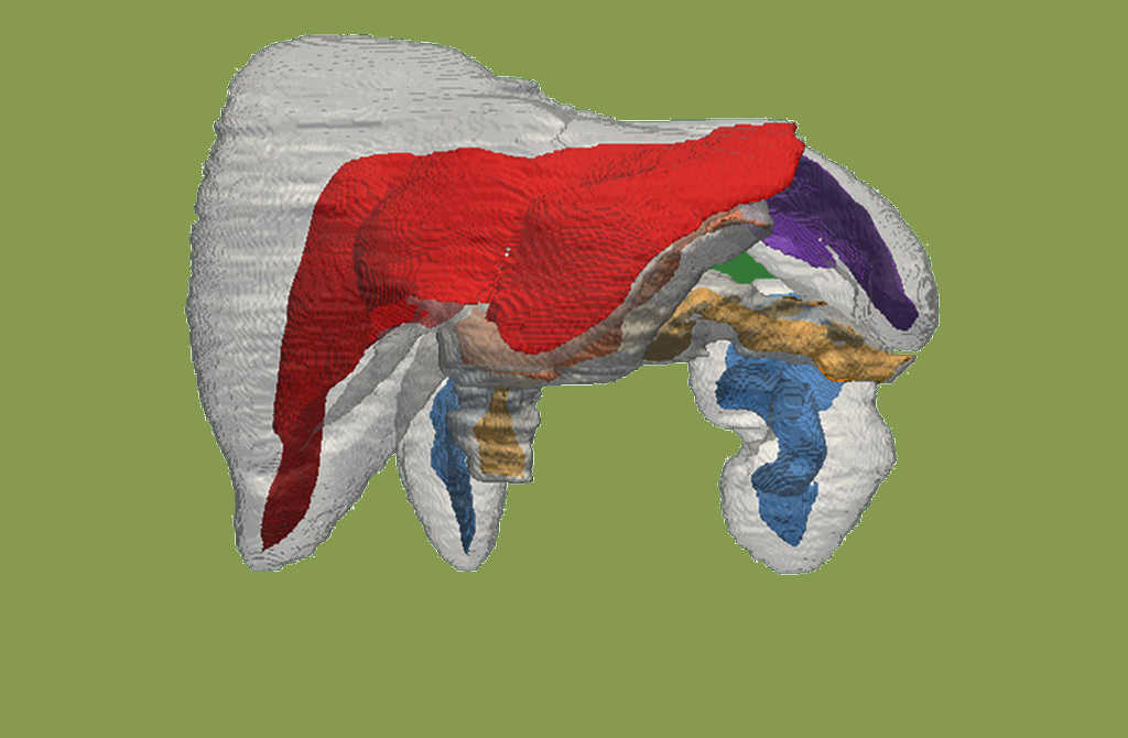
3D Bronchoscopy Navigation and Diagnosis system (3DEND) for increasing the yield of lung biopsy that incorporates a virtual bronchoscopy planner taking into account the bronchoscope’s kinematic constraints as well as bronchi breathing dynamics in order to guide the bronchoscope operator in real-time to peripheral pulmonary lesions that currently cannot be reached.
SAIPHIC
A system for automatic in-vivo polyp histology characterization (SAIPHIC) for providing the first on-line objective lesion diagnosis system considering both tissue visual features and an estimation of polyp size.